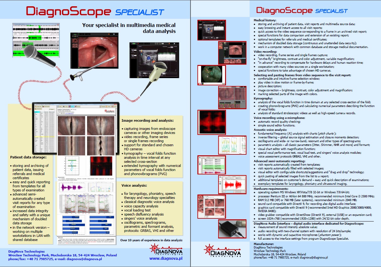
DiagnoScope – voice analysis and video recording environment
DiagnoScope (the previous version, for phoniatry only, was called DiagnoVoice) is a medical software that allows recording of endoscopic video sequences, and also in other medical specialties, where imaging devices are used for diagnostics, for example in ultrasound diagnostics. The version DiagnoScope Specialist (previously named DiagnoVoice Basic), designed for individual practitioners, is available since the beginning of 2010. The version DiagnoScope Clinic (previously named DiagnoVoice Lite) is designed for networked usage, e.g, in polyclinics.
Characteristics of DiagnoScope Specialist program:
- video recording, frame series or single frames recording,
- kymography,
- phonovibrogram (PVG),
- voice folds work parameters,
- dimensioning the object on video recording,
- brightness, contrast and color adjustment at each stage of video recording,
- additional recording settings with or without voice,
- reviewing source data at any time,
- automatic, configurable reporting,
- saving on any data-storage medium informations about report with all data,
- templates for specialist examinations,
- templates for referrals and medical certificates,
- modular construction allows individual configuration,
- optional modules on clients demand,
- data security by mechanism of double data entry,
- attendance of chosen HD cameras,
- multifunctional footswitch control,
- working in MS Windows 7, 8, 10, 11 environment,
- additional acoustic analysis module,
- optional examination templates on demand,
- possible to order installation and full configuration of program.
DiagnoScope Specialist modules:
- Video registration module allows to capture, edit and archiving pictures from endoscope camera or any other imaging device (ultrasound for example),
- Kymography is work analysis of the vocal folds as a function of time at any cross-section of the vocal folds, it allows to recognize defects in the vocal folds work (for example insufficiency of the vocal cords).
Features:
- Video recording recording is possible with an endoscope or another imaging device; available functions:
- video, frame series or single frames recording,
- “on-the-fly” brightness, contrast and color adjustment,
- variable magnification,
- recording “in advance” to compensate delays of human reaction and image processing and displaying by the computer,
- recording with or without sound (by setting);
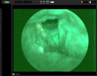
Example of video recording: an autofluorescence examination

The above video sequence with adjusted colors
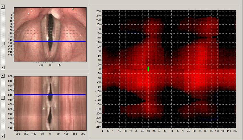
- Kymography module: work analysis of the vocal folds in time interval at any selected cross-section of the fold:
- an analysis based on recording using high-speed camera, line scan camera or stroboscope camera,
- convenient time interval selection window for kymographic analysis,
- intuitive mechanism to indicate the beginning and end of the vocal fold for analysis,
- time analysis of the vocal folds work in any cross-section of the vocal folds preview,
- adding vocal folds work frames to the report;
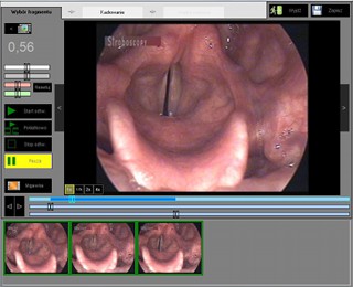
Video fragment selecting window for kymography
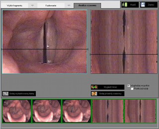
Time analysis of the vocal folds work preview window
- Phonovibrogram (PVG): visualization of entire vocal fold dynamics from slow motion video:
- slow motion video analysis,
- vocal fold dynamics quality assessment,
- image-processing algorithm that extracts the vocal fold motions of a whole laryngoscopic slow motion video film and compresses them into a single image,
- single image for full diagnosis;
Phonovibrogram visualization window; standard vocal folds work
Phonovibrogram visualization window; insufficiency of the vocal folds
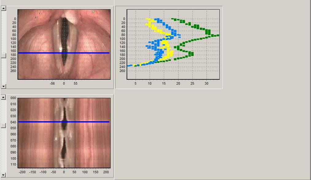
- Vocal folds work parameters: quantitative descriptions of vocal folds work:
- slow motion video analysis,
- informations about vocal folds work presented in the form of graphs:
- amplitude of movement of the vocal folds – evaluating the effectiveness of the work of the vocal folds,
- the ratio of time interval when glottis is open to time interval glottis is closed expressed in percent,
- the ratio of glottis midline motion amplitude to distance between glottis edges amplitude,
- presenting results of fitting glottis edges to determine difference between glottis motion phase,
- precise description of the degree of vocal fold insufficiency;
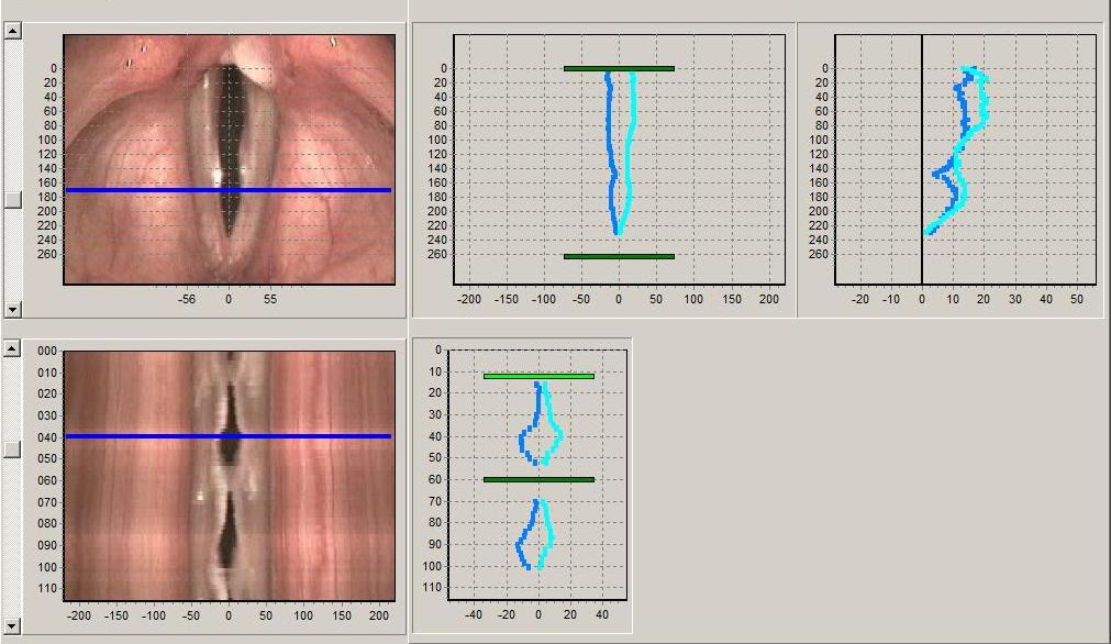
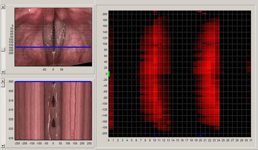
Vocal folds work parameters presentation window; glottis edges preview
Vocal folds work parameters presentation window; glottis motion amplitude
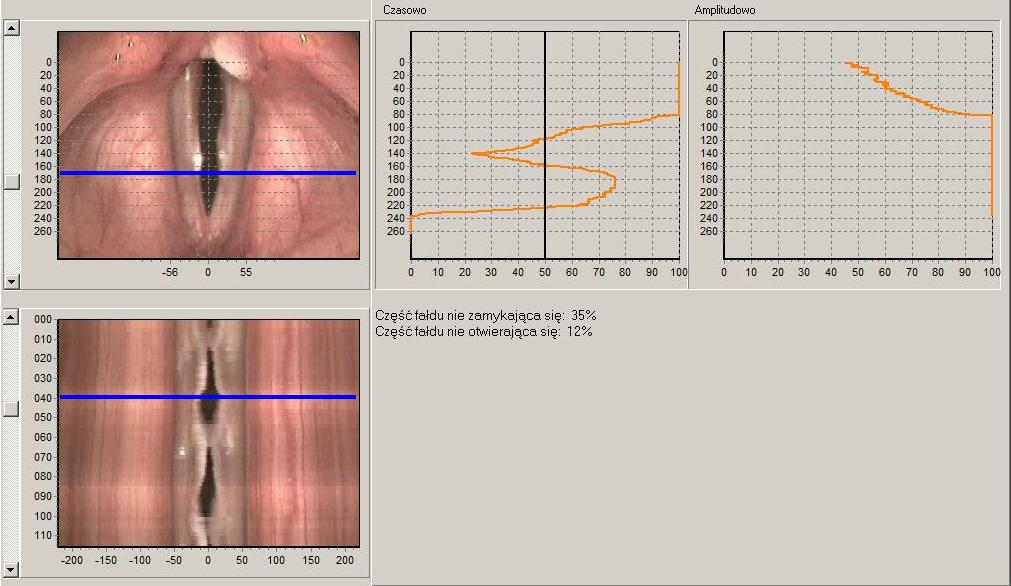
Vocal folds work parameters presentation window; glottis closure degree
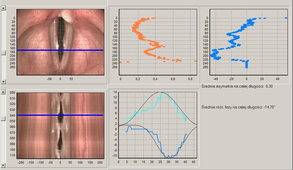
Vocal folds work parameters presentation window; vocal folds motion asymmetry
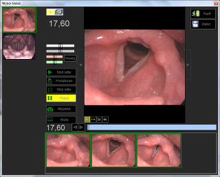
- Editing of video sequences: selecting frames from a video sequence allows collecting a set of pictures proper for diagnostics:
- comfortable and intuitive frame selection window (optional separated large frame selection window),
- possible to play video slow (frame by frame) makes selecting frames easier;
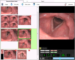
Frame selection window
- Dimensioning: measurement of length, width, perimeter and area of the selected object in the image
- determining the scale before measurement based on the object of known dimensions (e.g. ruler),
- easy object selecting using points,
- measurement of the object surface area,
- additional option of the length measurement in percentage;
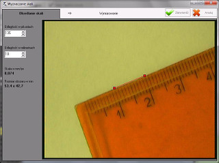
Determining the scale before measurement window

Dimensioning the objects window
- Review of source data: allows to quickly access the video sequence corresponding to a frame placed in an archived visit report;
- easy source data review widow of recorded video sequences, frame series and single frames,
- possible to play all recorded sequences any time;
Source data review window
- Editing of frames from a video sequence: at each report creating stage, allows adequate correction of images, ie
- brightness, contrast and color adjustment,
- magnification,
- marking selected parts of the image with colors;
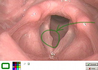
Marking selected parts of an image with image editor
- Advanced semi-automatic, configurable reporting:
- automatically created visit report forms from templates,
- automatically filled with selected images,
- editing in a visual editor,
- quick pasting of selected images from the list to a report;
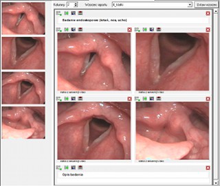
Automatically created and filled visit report
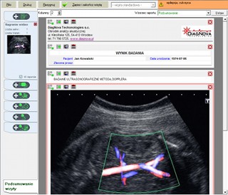
- Application for ultrasound diagnostics:
- optional templates created on clients demand makes examination description much easier and quicker,
- we have sample templates for ultrasound;
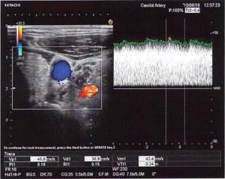
Example output of a frame from ultrasound Doppler examination
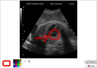
Picture from ultrasound with fragment marked with color
Report from ultrasound examination
- Application for gastrointestinal endoscopy:
- optional templates created on clients demand makes examination description much easier and quicker,
- capturing pictures with flexible endoscope with an endoscopic video channel(camera);
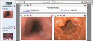
Exemplary report from gastrointestinal examination using endoscope
- Additional documents:
- optional templates created on clients demand for referrals and medical certificates,
- fast access from main window,
- easy template edition if needed;
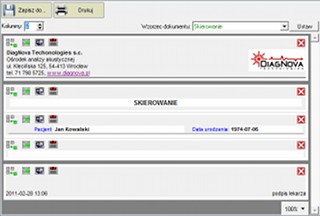
Window for issuing referrals and medical certificates
- Medical history:
- storing of patient data and visit reports,
- instant access,
- easy browsing,
- data comparison functions,
- complement of the existing report.
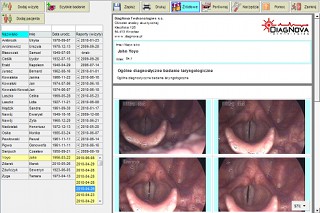
Main program window with patient data and visit reports
DiagnoScope in phoniatric and laryngologic application is available with additional voice analysis module.
Hardware requirements:
- Minimum requirements for video signal recording with resolution 352×288
- Processor: Intel Pentium III or later with clock frequency min. 800 MHz, AMD Athlon 64 or later with clock frequency min. 800 MHz;
- Sound card with DirectX 9 compatible drivers;
- Operating system: MS Windows XP 32 bit, MS Windows Vista 32 bit or later 32 bit;
- RAM at least 512 MB (for Windows XP) or at least 768 MB (Vista);
- Integrated graphics card compatible with DirectX 9;
- Hardware to convert analog video signal (frame grabber) compatible with DirectShow (DirectX 9), external (USB) or in the form of expansion card;
- 24 (32) bit color depth and screen resolution at least 1024×768 pixels.
- Minimum requirements for video recording signal with resolution 720x576
- Processor: at least dual-core with clock frequency min. 2000 MHz: Inte i3, AMD Ryzen 3;
- RAM at least 2048 MB (recommended 4096 MB, especially for kymography);
- Operating system: MS Windows 7/8/10 32-bit or 64-bit;
- Integrated graphics card compatible with DirectX 9;
- Hardware to convert analog video signal (frame grabber) compatible with DirectShow (DirectX 9), external (USB) or in the form of expansion card;
- 24 (32) bit color depth and screen resolution at least 1366×768 pixels.
- Minimum requirements for video recording signal with HD resolution 1280x720
- Processor: at least dual-core with clock frequency min. 2000 MHz: Inte i3, AMD Ryzen 3;
- RAM at least 4096 MB (recommended 8192 MB, especially for kymography);
- Operating system: MS Windows 7/8/10 64-bit;
- Integrated graphics card compatible with DirectX 9;
- Hardware to convert analog video signal (frame grabber) compatible with DirectShow (DirectX 9), external (USB 3.0) or in the form of expansion card;
- Two hard drives recommended: 240 GB SSD (for operating system) and 1 TB HDD (for files);
- 24 (32) bit color depth and screen resolution at least 1920×1080 pixels.
- Minimum requirements for video recording signal with FHD resolution 1920x1080
- Processor: at least 8-core or 6-core with 12 hardware thread with clock frequency min. 3500 MHz: Intel i7, AMD Ryzen 5;
- RAM at least 8192 MB;
- Operating system: MS Windows 7/8/10 64-bit;
- Integrated graphics card compatible with DirectX 9;
- Hardware to convert analog video signal (frame grabber) compatible with DirectShow (DirectX 9), external (USB 3.0) or in the form of expansion card;
- Two hard drives recommended: 240 GB SSD (for operating system) and 2 TB HDD (for files);
- 24 (32) bit color depth and screen resolution at least 1920×1080 pixels (recommended 1920x1200 pixels).
Additional information:
We invite you to learn more about the program's capabilities and its support. They are described in detail in the user's manual, which can be obtained free of charge in electronic form, by writing to the e-mail address serwis@diagnova.pl.
An advertising leaflet about the DiagnoScope Specialist program.